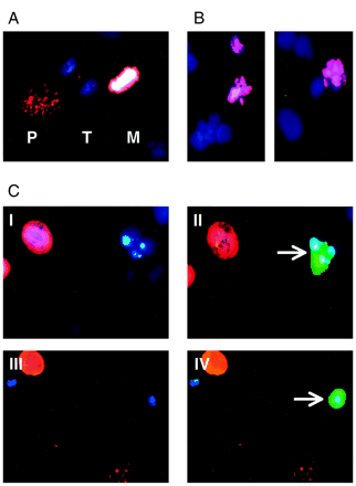
Fig. 5. Pin1 depletion induces apoptosis in interphase. A,
phosphorylation of histone H3 correlates with mitotic phases. Fixed
HeLa cells were double-stained with DAPI and with an antibody
recognizing phosphorylated histone H3. The nuclei of cells in prophase
(P), metaphase (M), and telophase
(T) are shown. B, mitotic cell death.
HeLa cells were incubated for 48 h with 50 nM Taxol
and double-stained as in A. C, Pin1
depletion induces apoptosis in interphase. Seventy-two h after
transfection with GFP and Pin1 antisense, fixed cells were
double-stained with DAPI and with an antibody recognizing
phosphorylated histone H3. II and IV,
same images as in I and III superimposed with GFP-emitted green
fluorescence. Arrows, transfected apoptotic cells.
